Digital Dental Imaging for Patients in Durham
Digital Dental Radiographs (x-rays)
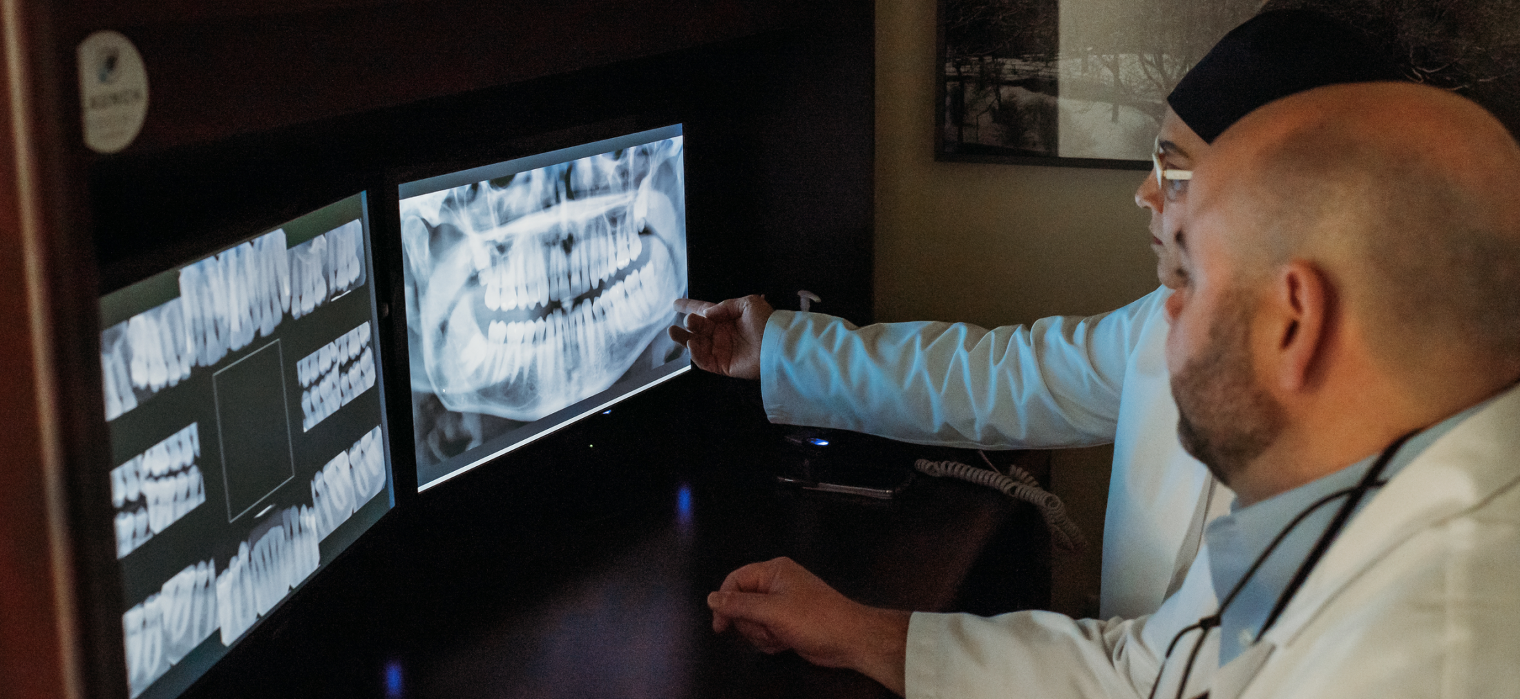
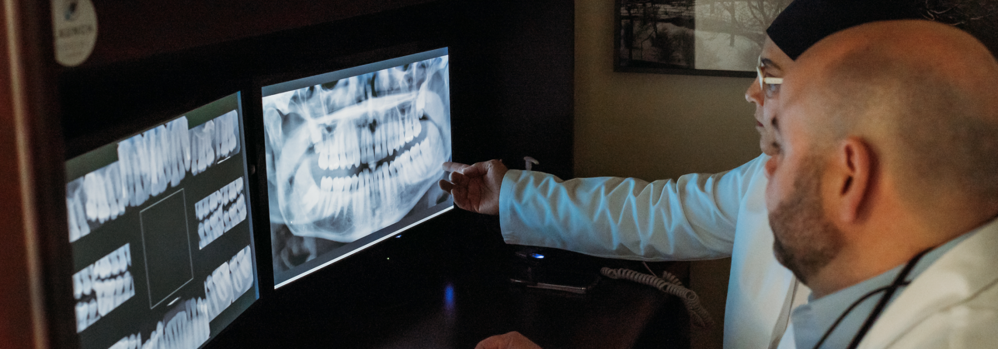
X-rays are invisible beams of ionizing radiation that are commonly and safely used both in medicine and in very low doses in dentistry. Once they pass through a body part, they are captured by our digital sensor and produce images (radiographs) in tones of gray, showing calcified structures such as the jaw bones, teeth, and other bony structures.
The use of digital x-rays to create dental images is not painful. The use of digital sensors has enabled dentists to much further reduce the radiation used compared to traditional film radiographs.
H&L Dentistry use digital radiographs to help diagnose disease that is not visible to the naked eye during a clinical dental examination, even with visual magnifications currently being used. As part of our preventive approach, we want to be able to detect any dental caries, anomaly, or pathology as early as possible to address it promptly.
How Safe Are Dental X-Rays?
How often X-rays should be taken depends on specific factors such as an individual’s current oral health, age, caries risk assessment, and any signs or symptoms of oral disease. This means that there is no “one size fits all” when it comes to the interval between dental X-rays.
Dentists adhere to the “ALARA” principle, a phrase established in 1973 by the International Commission on Radiologic Protection, that stands for “As Low as Reasonably Achievable.” Under the ALARA principle, dentists take precautions to help ensure that:
- X-rays are taken when needed and/or recommended.
- Necessary exposures are kept as low as reasonably achievable.
- The doses received by patients and personnel are kept well below the allowable limits.
Dental radiographs are proven to be safe. However, they do require very low levels of radiation exposure, which dramatically reduces the risk of a potential harmful effects. Dental radiographs account for approximately two and a half percent (2.5%) of the yearly effective dose received from other medical radiographs. Dental X-ray tools and techniques are designed to limit the body’s exposure to radiation and every precaution is taken to ensure that radiation exposure is As Low As Reasonable Achievable (the ALARA principle). A leaded apron minimizes exposure to the abdomen and may be used when it will not interfere with acquisition of the dental radiograph.
For more information about x-rays and digital imaging, visit the ADA’s website for X-ray information.
To schedule an appointment with Ivelis Hernández-Ramírez, DMD or Reinaldo Lasanta-García, DMD, please use the buttons below:

